Jun 5, 2020

Potential for Pressure injury ……

Pressure injury prevention (PIP) is an important area of patient safety. Encouraging patient participation in care is a growing trend in healthcare as it can increase adherence to treatment plans and improve outcomes.

Patients in acute care settings may be able to take on an active role in PIP. However, there is limited information on patients’ views of their perceived role in PIP. The aims of our study were to survey hospitalized patients’ views on

a) their perceived roles in PIP and,

b) factors that enable or inhibit patient participation in PIP strategies.


Pressure injuries (PIs) (also known as pressure sores, pressure ulcers or decubitus ulcers) are areas of localized damage affecting the skin and underlying tissue which results from pressure, and/or shear - Hospital acquired pressure injuries (HAPI).


Certain areas of our bodies are more prone to pressure sores, especially for individuals with limited mobility. For individuals confined to a wheelchair, the most common places for sores to occur are:

·         shoulder blades

·         spine or sacral area

·         back of the head

·         neck

·         back of the arms and legs

·          

For those that are typically bedridden, the most common places for pressure injuries are:

·         Hips

·         lower back

·         back of knees

·         back or sides of the head/ear


Each patient’s physical condition influences the potential for pressure injury:

·         general skin condition

·         nutritional status (weight, weight loss, serum albumin level)

·         fecal and urinary incontinence


There are several key recommendations to pressure injuries:

·         Avoid using hot water, and use only mild cleansing agents that minimize irritation and dryness of the skin. 

·         Avoid low humidity because it promotes scaling and dryness, which has been associated with pressure ulcer development.

·         During skincare, avoid vigorous massage over reddened, bony prominences because evidence suggests that this leads to deep tissue trauma.

·         Skin breakdown caused by friction may be mitigated by the use of lubricants, protective films, protective dressings, and protective padding.


To ensure successful participation in PIP, patients require education throughout admission, management of pain and discomfort and a supportive and collaborative relationship with health care staff.


Health professionals should identify patient ability and motivation to prevent pressure injury (PI), work in partnership with patients to adhere to PIP, and ensure that PIP actions are facilitated with appropriate pain relief.



PANCREATITIS


Definition :
Pancreatitis is an inflammation of the parenchyma of pancreas, an organ that is important in digestion.

Pathophysiology:
Auto digestion of the pancreas by its own proteolytic enzymes particularly trypsin leads to vasodilation, increase permeability, necrosis , erosion & hemorhhage leads to acute pancreatitis
Alcohol abuse & other etiologoical causes leads to inflammation & destruction of secreting cells  of pancrease causing maldigestion & malabsorption of protein & fat & these cells are then replaced by fibrous tissue causing obstructiuon of pancreatic & common bile duct leading to chronic panctreatitis.

Etiology & Risk factors :
Excessive alcohol consumption
Biliary tract diseases :
  • Cholelithiasis
  • Acute & Chronic Cholecystitis
Bacterial & Viral infection
Peptic ulcer
Ischemic Vascular Disease
Obstruction :
  • Cholidolithiasis
  • Sphincter of Oddi stenosis
  • Tumors
Trauma :
  • Blunt Abdominal trauma
  • Abdominal Operation
  • ERCP procedure
Metaboloc :
  • Acute hypercalcemia
  • Hypertrigylceridemia
Vascular :
  • Shock
  • Vasculitis
Toxins :
  • Alcohol
  • Snake & scorpion venom
Drugs:
  • Valproic acid,Azathioprine, Metronidazole, Tetracycline, ACE inhibitors


Types :
Acute Pancreatitis : Auto-digestion of pancreas by its own enzymes & development of fibrosis causing inflammation & edema to the pancreas. It may be self limiting or fatal.
Chronic Pancreatitis : Recurrent exacerbation leading to fibrosis after acute inflammation & persistent pancreatic tissue damage leading to decrease in pancreatic exocrine & endocrine function

Sign & symptoms :
  • Epigastric pain
  • Boring abdominal pain radiating to back or lest shoulder
  • Gnawing continous abdominal pain after intake of meal
  • Nausea & Vomiting (gastric origin but also bile stained)
  • Ascities
  • Weight loss
  • Blood glucose level fluctuation
  • Fatigue
  • Jaundice
  • Fever
  • Mental confusion
  • Tachycardia, cyanosis, cold, clammy skin & hypotension
  • Steatorrhea
  • Hypoglycemia, Hypocalcemia
  • DIC
  • DM

Medical & Nursing Management :
  • NPO status
  • IV Fluids for hydration
  • Vitamin Supplementation
  • Analgesics for pain management- IV/IM/Transdermal patch
  • H2 Receptor blockers : Rantidine & Cimetidine
  • Insulin therapy in case of DM or Hyperglycemia
  • Antipyretics & Antiemetics
  • Assess bilateral equality of lungs due to the presence of Ascites
  • Place patient in Knee chest position to relieve pain
  • Administer bland, low-fat, high protein, high calorie, small frequent meals after NG tube removal
  • Suction the NG tube after episodes of vomiting
  • Administer pancreatic enzymes
  • Monitor blood glucose levels

Distinctive Facts :
  • Ix: Elevated serum amylase, lipase, cholesterol, bilirubin, WBC, ERCP (Endoscopic Retrograde Cholangio Pancreatography )
  • Positive Turner's sign ( Bluish gray discoloration of flank areas) & Cullen's Sign ( Bluish gray discoloration of periumbilical area of abdomen)
  • Surgical Rx : Whipple procedure ( distal pancreatography), Autotransplantation of islets cells, Pancreaticojejunostomy, Drainage & Abrassion of pancreatic pseudocyst
  • Complications include Pleural effusion, GI hemorrhage,Shock, Pancreatic fistula, Necrosis & Abscess of Pancreas

Prognosis :
In the chronic form, episodes of pancreatitis tends to become more severe over time and the overall 10-year and 20-year survival rates are estimated to be about 70% and 45%, respectively.
Acute Pancreatitis may be life threatening if not treated


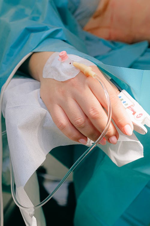
Patient with Iv Line


GUILLIAN-BARRE SYNDROME

                                                      GUILLIAN-BARRE SYNDROME


What is GBS?
 Guillain Barré Syndrome (GBS) is an autoimmune disease in which the peripheral nerves and nerve roots are damaged by the immune system’s antibodies and lymphocytes.The syndrome is named after the French neurologists Georges Guillain and Jean Alexandre Barré
Definition : 
It is a collection of clinical symptoms that manifests as an acute, frequently severe & fulminant polyradiculoneuropathy that is autoimmune in nature.

Pathophysiology :
Infectious organism containing amino acid that mimics the peripheral nerve myelin protien leads to cell mediatory inflammatory attack on peripheral nerve myelin as the immune system can not distinguish between the two leads to destruction of peripheral nerves targeting ganglioside GMB1 causing diminished nerve conduction.

Etiology & Risk factors :
  • Idiopathic
  • Viral Infection : Influenza, Zika virus, Epstein Barr Virus, Hepatits E virus etc
  • Bacterial infections: Mycoplasma pneumoniae, Campylobacter jejuni etc
  • Injury to spinal cord
  • Head Injury

Types :
  • Acute Inflammatory Demyeliunating Polyneuropathy [AIDP]: Inflammation & destruction of myelin sheaths of peripheral nerve cell activated by macrophages
  • Acute Motor Axonal Neuropathy [AMAN]:  Binding of antibodies to ganglioside antigens on axon cell membrane followed by macrophage invasion, inflammation & axonal damage.
  • Acute Motor & Sensory Axonal Neuropathy [AMSAN]: Bpoth motor & sensory nerve fibres are damaged due to macrophage invasion & inflammation causing axonal damage.
  • Miller Fisher Syndrome [MFS]:  Demyelination of nerve roots 

Sign & Symptoms :
Motor System : 
  • Motor paralysis with or without sensory disturbance
  • Ascending type paralysis
  • Rubbery legs
  • Dysesthesias of limbs
  • Lower limbs are affected more
  • Burning & Numbness or tickelin feeling on lower limbs
Deep Tendon Reflexes :
  • Attenuated or absent reflex
Cranial Nerves :
  • Facial diparesis
  • Bulbar weakness
  • Ophtalmoplegia
  • Pupillary paralysis
  • Optic atrophy
Bladder :
  • Loss of bladder tone & sensation is seen in early course of disease
Autoimmune Involvement :
  • More prominent
  • Labile BP
  • Cardiac dysrhythmias
  • Dyspnea due to intercoastal & diaphragmatic muscles are affected

Medical & Nursing Management :
  • Plasmapheresis is the treatment of choice
  • Corticosteroids to suppress immunity
  • Analgesics to reduce pain
  • Immunoglobulin Intravenously
  • Oxgenation
  • Turn & Reposition the patient
  • Assess Vital signs, rate of accessory muscle involvement
  • Assess communication ability
  • Symptomatic Mgt

Distinctive facts:
  • Ix- CSF analysis, Nerve Conduction Test, CT scan, MRI
  • Most common type is AIDP
  • GBS is also called as French Polio, Acute Idiopathic Polyneuritis
  • Complications are DVT, bed Sore, Pneumonia
  • Physiotherapy is mandatory

Prognosis:
Most of the patients recover fully but it takes many months of  intensive therapy. The mortality ranges from 2-12%. 15% patients suffer disability & 10% are unable to walk.


CARDIO-PULMONARY RESUSCITATION

                                                 CARDIOPULMONARY RESUSCITATION 


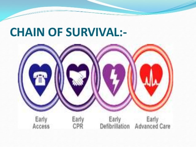
 CHAIN OF SURVIVAL:-

What is CPR?
CPR is a combination of rescue breathing (mouth-to-mouth resuscitation) and chest compressions to restore circulation of oxygen-rich blood to the brain. Without oxygen, permanent brain damage or death can occur in less than 8 minutes
Definition :
Cardiopulmonary resuscitation (CPR) is a combination of mouth-to-mouth resuscitation and chest compressions that delivers oxygen and artificial blood circulation to a person who is in cardiac arrest. It can be life-saving first aid.
Indications of CPR:
Cardiac Arrest
  •        Heart disease – the most common cause of cardiac arrest
  •       Ventricular fibrillation (VF)
  •       Ventricular tachycardia (VT)
  •        Asystole
  •        Pulse less electrical activity
Respiratory Arresst
THIS CAN BE DUE TO FOLLOWING:
  •       Drowning
  •       Stroke
  •       Foreign body in throat
  •       Smoke inhalation
  •       Drug overdose
  •       Electrocution or injury by lightening
  •       Suffocation
  •       Accident, injury
  •       Coma
  •       Epiglottis paralysis.
  Metabolic Indications : 
  •       Hypoglcemia
  •       Electrolyte Imbalane
        Fluid Balanace:
  •       Hypovolemic Shock
  •       Hemorrhage
       Neurologic Indications :
  •       Massive CVA
       Poisons or Substance Overdose:
  •       CO Poisoning
  •       Proipanolol Overdos
Purpose of CPR:
  •      To maintain an open and clear airway (A).
  •      To maintain breathing by external ventilation (B).
  •      To maintain Blood circulation by external cardiac massages (C).
  •      To save life of the Patient.
  •      To provide basic life support till medical and advanced life support arrives

Warning Sings of Cardio Pulmonary Arrest:
  •       Loss of consciousness
  •      Convulsions
  •      Apnea or stridor
  •      Dilated pupils
  •      Absence of heart rate or weak thready pulse
  •      Cyanosis
  •      Hypothermia
Procedure of CPR:
Check for response by tapping on shoulder & saying "HEY ARE YOU OK?''
Airway :
  •      Head Tilt Jaw Left Manoeuvre :While pushing the forehead back tilt the chin forward
  •      Jaw Thrust Manoeuvre : This is to be performed while neck injury by pushing the face forward with index finger by holding cheeks with both the hands 
Breathing : Look, Listen & Feel for breathing & pulse. Breathing can be initiated by :
  •      Mouth to Mouth Ventilation
  •      Mouth to Mask Ventilation
  •      Bag Mask Ventilation
Circulation : Check for pulse:
  •      Adult : Carotid artery
  •      Infant : Brachial artery
Chest Compressions :
Interlock fingers of both the hand by placing heel of domninant hand on the centre of chest & other hand on its top.
For infants : Place 2 fingers or 2 thumbs on the breast bone & give downward thrust by tilting the head back of infant
Universal Compression to Ventilation Ratio:
  •       For Adult - 30:2
  •       For Infant & Children - 15:2
  •       For Newborn - 3:1
Recommended Depth of Compression:
  •      In adult & children- 5 cm [2 inches]
  •      In infants - 4 cm [1.5 inches]
Rate of Compression = 100/min

Complications of CPR:
  •      Rib & Spleen fracture
  •      Pneumothorax & Hemothorax
  •      Injury to diaphragm, Myocardial muscle
  •      Aspiration
  •      Vomiting

Medical & Nursing Management during CPR
  •       Adrenaline: to enhance cerebral and myocardial blood flow by preventing arterial collapse
  •       Noradrenaline : to restore normal circulation
  •       Atropine: to block vagal tone completely & enhances automacticity and conduction of both sinoatrial and atriventricular node.
  •       Amiodarone or Lidocaine : to dilate coronary artery  leading to vasodilatation and reduction in afterload and systemic blood pressure.
  •       Calcium Gluconate :enhances the contractile force of cardiac muscle
  •       Sodium Bicarboinate :to alkalinaze urine in drug over dose
  •       Magnesium : to stabilize myocardial cell membrane
  •       Intubation & Oxygenation
  •       Administer IV fluids
  •       Documentation of Vitals & urine output
  •       Maintain appropriate ventilation


Jun 2, 2020

MYOCARDIAL INFARCTION

                                                          MYOCARDIAL INFARCTION


What is MI?
MI refers to the process by which areas of the myocardial cells in the heart are permanently destroyed.

Definition :
MI is a disease condition which is caused by reduced blood flow in the coronary artery due to atherosclerosis & occlusion of the artery by embolus or thrombus.

Pathophysiology :
Causative factors will lead to atherosclerosis causing inadequate oxygen supply to myocardial cells due to narrowing of coronary arteries followed by cell death causing inflammation & release of cardiac enzymes leading to accumulation of lactic acid due to anerobic glycolysis leading to infarction & chest pain.

Etiology & Risk factors :
Modifiable :
  • High blood pressure 
  • Stress
  • High blood cholesterol
  • Obesity
  • Smoking & Alcoholism
  • Sedentary lifestyle
  • Diabetes Mellitus
Non modifiable :
  • Age : more than 40 years
  • Gender:  Male:Female = 3:1
  • Family history : Inherited to children from parents


Types of MI:
Transmural Infarction : Extension of infarcts through the whole  thickness of heart muscle & are usually a result of complete occlusion in the area's blood supply. It is also called as ST Elevation MI [STEMI].
Subendocardial Infarction : Involves the small area in the endocardial wall of left atrium, left ventricles or papillary muscles. It is also called as Non transmural or Non ST Elevation MI [NSTEMI]


Sign & Symptoms :
  • Chest pain unrelieved by rest or NTG
  • Pain that radiates to arms, jaw, back or neck
  • Dyspnea
  • Nausea & Vomiting
  • Tachycardia > 100 beats/min
  • Variable blood pressure
  • Anxiety
  • Restlessness
  • Diaphoresis
  • Pale, Cool, Clammy skin
  • Death

Medical & Nursing Management :
  • Administer oxygen & Aspirin
  • AntiArhythmics : Amiodaron
  • Antihypertensive to keep BP low ; Hydralazine
  • Thrombolytics to reestablish blood flow in occluded artery : streptokinase
  • Heparin following thrombolytic therapy
  • Calcium channel blockers to prevent reinfarction & ischemia : Verapamil
  • Beta adrenergic blockers to reduce duration of ischemic pain & ventricular fibrillation : Propanolol, nadolol, metoprolol
  • Analgesics to relieve pain : Morphine
  • Nitrates to dilate blood vessels : Nitroglycerine
  • Note foe PQRST [Precipitating events, Quality of pain, Radiation of pain, severity of pain, Timing]
  • Electrical Cardioversion
  • Assess heart sounds & heart murmur
  • Assess Cardinal Signs
  • Educate patients for lifestyle mofification

Distinctive facts :
  • Ix- ECG,Cardiac enzymes :  CK-MB elevated, Troponin-I & Troponin T elevates within 1 hour of MI
  • T- wave inversion : Ischemia
  • ST elevation/depression : Injury
  • Significant Q waves : Infarction
  • Also called as Heart Attack
  • Silent MI more common in diabetic patient
  • Accronym for treatment - MONAT Therapy [Morphine,Oxygen,Nitroglycerine, Aspirin, Thrombolytics]
  • Complications include death
  • Surgical Rx- PTCA, Stenting, CABG, Athrectomy,
  • CPR in case of arrest
  • Obstruction of LAD results in Anterior or Septal wall MI
  • Obstruction of Circumflex artery results in Posterior or Anterior wall MI
  • Obstruction of Right Coronary Artery results in Inferior wall MI 

Prognosis :
Prognosis is good in patients receiving appropriate medical attention & lifestyle modification as early as possible. Mortality is however high among patients of MI


Jun 1, 2020

HYPERTENSION

                                                                      HYPERTENSION 
Definition:
Hypertension is defined as the sustained increase in blood pressure more than 140/90 mm hg on average 2 times of sitting reading.

Pathophysiology :
Etiological factors will cause activation of vasoactive substance leading to vasoconstriction in peripheral arterioles causing activation of renin charecterized by Angiotensin II release leading to sodium & water retention resulting in high blood pressure

Etiology & risk factors :
  • Primary Hypertension : elevation in BP without identified cause
  • Secondary Hypertension : elevation of BP with exact cause
Causes of Secondary Hypertension:
  • Congenital Narrowing of Aorta
  • Renal disease
  • Endocrine disorders like Cushing's syndrome
  • Sleep Apnea
  • Brain disorders including brain tumor or injury
  • Medications like NSAIDs, cocaine
  • Liver cirrhosis
Risk factors :
  • Family history
  • Genetics
  • Obesity
  • Age over 50 years
  • Sedentary lifestyle
  • Stress
  • Alcoholism, Smoking

Types of HTN:
  • Pre HTN :SBP- 120-139 mmHG  DSB80-89 mmHG
  • Hypertension stage I - SBP140-159 mmHG   DBP-90-99 mmHg
  • Hypertension Stage II - SBP: More or equal to 160 mmHg  DSB :More or equal to 100 mmHG
  • Pregnancy induced Hypertension : Rise in BP more tha 140/90 mm Hg during pregnancy & subsides after 12 weeks of post partum 

Sign & Symptoms :
  • Asymptomatic 
  • Headache 
  • Dizziness
  • Blurred vision 
  • Nausea/ vomiting
  • Epistaxis
  • Chest pain
  • Shortness of breath
  • Pappiledema

Medical & Nursing Management :
Step I : Non Pharmacological Measures :
  • DASH diet
  • No smokin, alcohol & caffeine reduction
Step II : Pharmac Measures
  • Diuretics - Furosemide, Spironolactone, Bumetanide
  • Beta Adrenergic Blockers to block agents to lower HR & Cardiac output - Propanolol, Atenolol, Metoprolol
  • Calcium Channel Blockers to cause peripheral vasodilation - Nifedipine, Necardipine
  • ACE inhinitors to inhibit Renin Angiotensin Aldosterone System-  Enalpril, Captopril
Step III- Increase dosages of currently administered medication
Step IV- Combination of multiple drugs

Distinctive Facts:
  •  Ix- BP monitoring, EKG, BUN, Lipid Profile
  • Also called as "SILENT KILLER "
  • Complications include Stroke, Retinal Damage, Heart failure
  • Identification of "WHITE COAT" Hypertension is mandatory
  • Benign Hypertension distinguishes this type of high blood pressure from the more aggressive and rapidly developing accelerated hypertension which is also known as malignant hypertension.
  • Malignant high blood pressure is more acute and severe rapidly reaching its end stage which may be stroke, heart attack or heart failure if left unattended.


Prognosis :
  • Majority patients have primary HTN & patients are asymptomatic. 
  • Life style modifications & medicine intake are must to reduce end organ damage to vital organs.


110 - Nursing Exams Questions & Answers - Svastham Exemplar

  Question 5476) Which factor would most likely be a cause of epiglottitis?  A. Acquiring the child’s first puppy the day before the onset o...